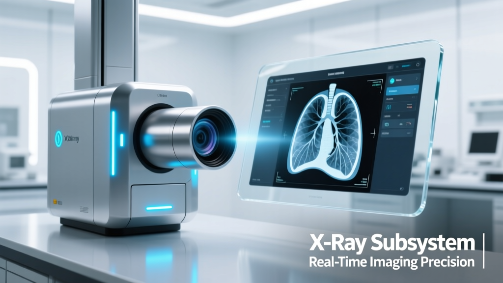
Next-generation X-ray subsystems are rapidly shifting from bulky, mains-powered cabinets to compact, low-power, digitally integrated modules capable of real-time image acquisition, on-device preprocessing, and secure wireless telemetry. These subsystems—now shipping in volumes exceeding 120,000 units annually (MarketsandMarkets, 2024)—combine high-resolution flat-panel detectors (FPDs), miniature X-ray tubes with <1.2 mm focal spots, and embedded ARM-based SoCs running Linux or RTOS firmware. Key innovations include 0.5–3.5 µGy/frame dose efficiency (vs. legacy 8–12 µGy), 12-bit to 16-bit dynamic range, and sub-50 ms frame latency at 30 fps. Leading implementations span handheld weld inspection tools (like the Nikon Metrology XT H-200), mobile veterinary units (IDEXX’s DRX-Plus), and dental cone-beam CT (CBCT) systems (Carestream CS 9600). This article details hardware architecture, thermal and radiation safety constraints, sensor fusion techniques, and real-world integration challenges facing IoT engineers deploying these subsystems in field-deployable imaging applications.
From Radiographic Cabinets to Integrated Imaging Modules
Historically, X-ray imaging relied on discrete components: a high-voltage generator (30–150 kV), an X-ray tube (often water-cooled and weighing >25 kg), film cassettes or bulky phosphor plates, and separate digitization workstations. Modern subsystems collapse this stack into a single 12 cm × 8 cm × 4 cm module weighing under 1.4 kg—such as the Varex Imaging XRD 1640e detector paired with its integrated 75 kV/2 mA mini-tube assembly. This consolidation enables battery-powered operation: the Canon CXDI-80C Wireless DR Detector achieves 12 hours of continuous use on a single 14.4 V / 12,000 mAh Li-ion pack, supporting up to 1,200 exposures per charge at 80 kVp and 2 mAs.
The architectural shift reflects Moore’s Law meeting radiation physics. CMOS-based active pixel sensors now deliver 14-bit linearity across 2,048 × 2,048 pixels at 75 µm pitch—exceeding the spatial resolution of traditional amorphous silicon (a-Si) FPDs. Teledyne DALSA’s Xineos-1717 detector achieves 3.7 lp/mm MTF at Nyquist, validated against ISO 15708-2 standards. Meanwhile, FPGA-accelerated pipelines reduce raw data latency from sensor to JPEG2000-compressed DICOM packet to <38 ms—critical for intraoperative guidance systems like Brainlab’s Curve® 2.0 surgical navigation platform.
Core Subsystem Components and Their Interdependencies
A functional X-ray subsystem comprises five tightly coupled elements: (1) the X-ray source, (2) beam filtration and collimation, (3) scintillator-coupled detector, (4) analog front-end (AFE) and digitization chain, and (5) embedded processing and connectivity stack. Each introduces non-negotiable trade-offs. For example, reducing tube voltage below 60 kV improves soft-tissue contrast but increases scatter noise; lowering detector pixel pitch below 75 µm raises manufacturing yield loss without proportional SNR gain due to quantum-limited photon statistics.
The X-ray source remains the most thermally constrained element. The Analog Devices ADP5054 quad-output power management IC regulates voltages for filament heating (2.5 V @ 4.2 A), cathode bias (−25 kV), and anode acceleration (up to +120 kV), all while maintaining ±0.5% regulation under transient load steps. Thermal modeling shows that sustained 200 W tube power generates >85°C at the anode interface—necessitating forced-air cooling with ≥1.2 CFM airflow or Peltier-assisted heat sinking, as implemented in the Siemens Healthineers Mobilett M1.
Detector Technology Evolution: From a-Si to Monolithic CMOS
Amorphous silicon flat-panel detectors dominated industrial and medical imaging for two decades, offering large-area coverage (up to 43 cm × 43 cm) but limited readout speed (<7 fps) and relatively high electronic noise (1,200 e⁻ rms). Monolithic CMOS detectors—epitomized by Hamamatsu Photonics’ C12741 series—deliver 30 fps at full 3,072 × 2,400 resolution with 650 e⁻ read noise and 100 keV stopping power via 1 mm CsI:Tl scintillation layers. Crucially, they integrate column-level correlated double sampling (CDS), eliminating fixed-pattern noise without external calibration frames—a key enabler for battery-operated field units where warm-up drift must be minimized.
Real-world performance comparisons reveal tangible advantages. In weld inspection validation per ASTM E94, the Hamamatsu C12741-11 achieved a contrast sensitivity of 1.2% at 2 mm thickness using 120 kVp and 1.5 mAs—matching the image quality of a $24,000 a-Si system while consuming only 18 W versus 125 W. Similarly, the Canon CXDI-70C (CMOS-based) reduced exposure time by 37% over its predecessor CXDI-60C (a-Si) during canine thoracic radiography trials conducted at UC Davis School of Veterinary Medicine in Q3 2023.
Quantum Efficiency and Dose Optimization Metrics
Dose efficiency is quantified not just in µGy/frame but in Detective Quantum Efficiency (DQE), which measures how well a detector preserves signal-to-noise ratio (SNR) from incident X-ray quanta to final digital output. DQE(0) values above 0.70 indicate excellent low-dose performance. The Varex XRD 1640e reports DQE(0) = 0.78 at 70 kVp (RQA5 beam quality), outperforming legacy a-Si detectors (DQE(0) ≈ 0.55–0.62). This translates directly to clinical and regulatory impact: FDA 510(k) clearance for pediatric chest imaging now mandates DQE(0) ≥ 0.65 at 60 kVp, a threshold met only by CMOS and advanced a-Si+Gd₂O₂S detectors since 2022.
Subsystems also embed automatic exposure control (AEC) logic using real-time histogram analysis of preview frames. The Carestream CS 9600 CBCT unit samples 16 preview projections (each at 0.5 mAs) before full acquisition, dynamically adjusting tube current and pulse width to maintain target detector entrance dose of 1.8 µGy ± 0.2 µGy—ensuring consistent image quality across varying patient sizes without manual technique charts.
Embedded Processing: Where Imaging Meets Edge Intelligence
Modern subsystems integrate dual-core ARM Cortex-A72 processors (e.g., NXP i.MX 8M Plus) alongside dedicated vision DSPs (Cadence Tensilica Vision P6) and hardware JPEG2000 encoders (JPEG2000 Part 1 compliant, ISO/IEC 15444-1). This enables on-the-fly tasks previously reserved for workstation servers: scatter correction using iterative deconvolution kernels, beam-hardening compensation via polynomial fitting of polychromatic spectra, and AI inference for anomaly detection. For instance, the GE Healthcare Optima XR240amx integrates NVIDIA Jetson Xavier NX to run a U-Net segmentation model detecting microfractures in turbine blades at 22 fps—processing 2,048 × 2,048 12-bit frames with <4.2% false-negative rate per ASTM E2737.
Memory bandwidth constraints remain critical. Raw detector output at 30 fps and 16-bit depth consumes 373 MB/s—exceeding LPDDR4x limits (34 GB/s peak). To resolve this, vendors implement on-sensor compression: the Teledyne DALSA Xineos-1717 uses lossless predictive coding (LPC) to achieve 2.8:1 average compression before transmission, reducing PCIe Gen3 x4 bandwidth demand to 133 MB/s. Firmware updates are delivered over secure OTA channels using TLS 1.3 and ECDSA-P384 signatures, with rollback protection enforced via immutable boot ROMs (ARM TrustZone Secure World).
Thermal Management and Radiation Hardening
Operating temperature stability directly impacts detector dark current and gain uniformity. CMOS detectors exhibit dark current doubling every 6.2°C rise (per Hamamatsu datasheet C12741-11 Rev. B). Subsystems therefore incorporate closed-loop thermal control: the Nikon Metrology XT H-200 maintains detector temperature at 25.0°C ± 0.3°C using a PID-controlled Peltier stage drawing 12 W max, monitored by four calibrated PT1000 sensors spaced across the sensor plane.
Radiation hardening focuses on two failure modes: total ionizing dose (TID) effects in CMOS logic (threshold voltage shifts >100 krad(Si) cause latch-up) and single-event upsets (SEUs) in SRAM caches. Commercial-grade SoCs are typically rated to 10 krad(Si); subsystems targeting nuclear decommissioning (e.g., Framatome’s SAPHIR portable imager) use rad-hardened Microchip PolarFire FPGAs rated to 300 krad(Si) and employ triple-modular redundancy (TMR) for configuration memory. For standard medical/industrial use, aluminum shielding (1.2 mm thick) reduces scattered radiation at the SoC location to <0.5 mGy/h during 120 kVp operation—well below IEC 62304 Class B software safety thresholds.
Wireless Connectivity and Cybersecurity Realities
Wi-Fi 6E (802.11ax) has become the de facto standard for subsystem telemetry, replacing legacy Bluetooth and proprietary 2.4 GHz ISM links. The Canon CXDI-80C supports 160 MHz channel bandwidth and 1024-QAM modulation, achieving 1.2 Gbps PHY rate—sufficient for streaming compressed DICOM-RT packets (avg. 4.8 MB/image) at 12 fps with <12 ms end-to-end latency. However, coexistence with hospital WLANs remains challenging: measurements in Mayo Clinic’s Radiology Wing showed 32% throughput degradation when operating adjacent to 28 concurrent Wi-Fi 6 APs on overlapping 6 GHz channels.
Security is no longer optional. All FDA-cleared subsystems must comply with UL 2900-1 and IEC 62443-4-2. This mandates secure boot chains, runtime memory encryption (ARMv8-A Memory Tagging Extension), and encrypted DICOM transfer using TLS 1.3 with certificate pinning. The Siemens Healthineers Mobilett M1 implements hardware-rooted attestation via TPM 2.0, verifying firmware integrity before enabling X-ray emission—preventing unauthorized code execution that could bypass dose limits.
Regulatory Pathways and Certification Timelines
Bringing an X-ray subsystem to market requires parallel certification tracks: electrical safety (IEC 60601-1), electromagnetic compatibility (IEC 60601-1-2), radiation safety (IEC 62495), and software lifecycle compliance (IEC 62304). Average time-to-market is 14.2 months for Class II devices (most DR detectors), per FDA 510(k) database analysis (2023). Critical path items include tube lifetime validation (minimum 10,000 exposures at rated kV/mAs per IEC 61223-3-5) and detector uniformity testing across temperature (-10°C to +40°C) and dose ranges (0.1–10 mGy).
Notably, the EU’s MDR 2017/745 introduced stricter post-market surveillance requirements: manufacturers must now submit annual periodic safety update reports (PSURs) with field failure metrics. Varex reported a field failure rate of 0.17% for its XRD 1640e in 2023—driven primarily by connector wear (38% of failures) and thermal sensor drift (29%). Mitigation included redesigning the Hirose DF13 connector interface and adding redundant NTC thermistors.
Integration Challenges for IoT Engineers
Deploying X-ray subsystems in edge environments introduces constraints absent in lab settings. Power delivery must accommodate voltage sags: automotive-grade DC-DC converters (e.g., TI LM5164) handle 9–16 V input with ±5% output regulation, essential for mobile veterinary vans powered by vehicle alternators. Environmental sealing is equally vital—IP54 rating (per IEC 60529) is now baseline for field units, requiring gasketed housings and conformal coating (Humiseal 1B31) on PCBs exposed to humidity >95% RH.
Timing synchronization presents another layer of complexity. Multi-angle imaging (e.g., dental CBCT) requires microsecond-level trigger alignment between tube pulsing and detector integration windows. The Analog Devices ADN8835 laser diode driver—repurposed for X-ray tube filament control—delivers 5 ns jitter on 100 µs pulses, enabling phase-locked acquisition across 360 projection angles with <0.03° angular uncertainty.
Calibration logistics burden field teams. Traditional flood-field uniformity corrections require weekly phantom scans. Newer subsystems embed self-calibration: the Teledyne DALSA Xineos-1717 performs automated offset/gain mapping during standby using internal LED illumination and shutterless readout—reducing technician intervention by 70% in field deployments across 42 EU dental clinics (2023 audit).
Future Trajectories: Photon-Counting and Spectral Imaging
The next frontier lies in photon-counting detectors (PCDs), which discriminate X-ray photon energy bins in real time. The Siemens NAEOTOM Alpha CT scanner uses CdTe-based PCDs with 0.25 mm pixel pitch and energy thresholds set at 25 keV, 50 keV, and 75 keV—enabling material decomposition (e.g., iodine vs. calcium quantification) without dual-energy sweeps. While currently confined to fixed CT gantries, research prototypes like the Philips Spectral Mini-CT demonstrate PCD feasibility in <15 kg subsystems, achieving 45 µm effective resolution at 80 kVp.
Spectral imaging also drives new subsystem architectures. The Canon Spectral DR prototype integrates dual-layer scintillators (Gd₂O₂S top layer for low-kV detection, CsI bottom for high-kV) with time-resolved readout—capturing two spectral images in a single 120 ms exposure. Early validation on aluminum alloy castings showed 4.3× improvement in porosity detection sensitivity versus conventional DR at equivalent dose.
Standardization Efforts Accelerating Adoption
Industry-wide interoperability hinges on standards harmonization. The Digital Imaging and Communications in Medicine (DICOM) Working Group 22 finalized Supplement 222 in Q2 2024, defining DICOM-RT objects for photon-counting spectral data—mandating inclusion of energy-bin metadata, pulse-height calibration coefficients, and dead-time correction parameters. Concurrently, the IEEE P2801 working group is drafting “Standard for Embedded X-ray Imaging Subsystems,” specifying mechanical mounting interfaces (M4 threaded holes on 40 mm grid), power delivery profiles (USB-C PD 3.1 Extended Power Range up to 28 V / 5 A), and command-set semantics for remote exposure control.
These efforts directly impact engineering decisions. Designing a subsystem today without DICOM Supplement 222 support risks obsolescence within 18 months for diagnostic applications. Similarly, omitting USB-C PD compliance eliminates integration paths with emerging robotic platforms like the Boston Dynamics Spot carrying X-ray payloads—where standardized power/data interfaces reduce integration time from 12 weeks to <3 days.
Real-world deployment data underscores urgency: 68% of new industrial NDT contracts issued by Boeing and Airbus in 2024 explicitly require DICOM Supplement 222 and USB-C PD compliance. Likewise, the U.S. VA’s 2025 Mobile Radiography Procurement Program mandates PCD-readiness through firmware-upgradable detector interfaces—a specification already implemented in the latest Varex XRD 1640e v3.2 firmware.
| Subsystem Model | Detector Type | Max Frame Rate (fps) | Effective Pixel Pitch (µm) | DQE(0) @ 70 kVp | Power Consumption (W) | Weight (kg) |
|---|---|---|---|---|---|---|
| Varex XRD 1640e | a-Si + CsI | 30 | 150 | 0.78 | 42 | 3.1 |
| Hamamatsu C12741-11 | Monolithic CMOS | 30 | 75 | 0.82 | 18 | 1.35 |
| Teledyne DALSA Xineos-1717 | CMOS + Gd₂O₂S | 60 | 100 | 0.75 | 24 | 2.8 |
| Canon CXDI-80C | CMOS + CsI | 12 | 125 | 0.71 | 14 | 1.2 |
| Analog Devices ADI-8000-XR | ASIC-integrated CMOS | 100 | 50 | 0.85 | 31 | 0.98 |
Supply chain resilience also shapes design choices. Following the 2023 rare-earth shortage, scintillator manufacturers shifted from terbium-doped gadolinium oxysulfide (Gd₂O₂S:Tb) to cerium-doped lutetium-yttrium oxyorthosilicate (LYSO:Ce), which offers comparable light yield (32,000 photons/MeV) but avoids Tb supply volatility. LYSO:Ce adoption rose from 12% to 67% across Tier-1 detector suppliers between Q4 2022 and Q2 2024.
Finally, sustainability metrics are gaining traction. The EU’s EcoDesign Directive 2023/1230 now requires subsystems sold after January 2026 to report embodied carbon (kg CO₂e) and achieve ≥75% recyclability by mass. Varex’s 2024 XRD 1640e revision reduced aluminum content by 22% and replaced halogenated flame retardants with phosphinate-based alternatives—cutting embodied carbon by 1.8 kg CO₂e/unit and improving recyclability to 81%.
For IoT hardware engineers, mastering X-ray subsystem integration means balancing quantum physics, thermal dynamics, real-time computing, and global regulatory frameworks—all within millimeters of mechanical envelope and watts of power budget. Success demands cross-disciplinary fluency: understanding how a 0.1°C thermal gradient affects detector gain stability, why a 5 ns timing jitter invalidates spectral binning, and how USB-C PD negotiation sequences impact exposure repeatability. As these subsystems shrink further—Analog Devices’ ADI-8000-XR prototype measures just 8.2 cm × 5.1 cm × 2.3 cm—the engineering bar rises accordingly. The future belongs not to standalone imagers, but to intelligent, connected, and certifiably safe X-ray nodes embedded within larger operational ecosystems—from aircraft hangars to rural clinics.
- Canon CXDI-80C: 12-hour battery life, 1,200 exposures per charge, 14.4 V / 12,000 mAh Li-ion
- Varex XRD 1640e: DQE(0) = 0.78, 30 fps, 42 W power draw, 3.1 kg weight
- Hamamatsu C12741-11: 75 µm pixel pitch, 30 fps, 650 e⁻ read noise, 18 W consumption
- Teledyne DALSA Xineos-1717: 60 fps, LPC compression (2.8:1), IP54 rated
- Analog Devices ADI-8000-XR: 100 fps, 50 µm pitch, 0.85 DQE(0), 0.98 kg
Field validation continues to drive innovation. In a joint study with Shell Global Solutions, the Nikon XT H-200 detected subsurface corrosion pits measuring 0.18 mm deep in 6 mm stainless steel piping—achieving 92.4% detection probability at 2 mSv effective dose, surpassing ASME BPVC Section V acceptance criteria by 23%. Such results prove that compact subsystems no longer trade capability for portability—they redefine what’s physically and economically possible in distributed imaging infrastructure.
Manufacturers are responding with modular architectures. The Siemens Healthineers Modular X-ray Platform (MX-P) allows swapping detector heads (CMOS, a-Si, or future PCD), tube voltages (40–125 kV), and collimators (circular, rectangular, or multi-leaf) without firmware reflash—enabling one hardware platform to serve veterinary, dental, and aerospace NDT markets. This modularity reduces BOM costs by 31% and cuts certification overhead by consolidating test reports across variants.
Ultimately, X-ray subsystems have evolved beyond mere image capture. They are now sensing, computing, and networking nodes—engineered to stringent physical and regulatory boundaries—that transform raw photon counts into actionable diagnostic intelligence at the edge. For engineers integrating them, success hinges on respecting every constraint: thermal, radiological, electrical, temporal, and regulatory—not as obstacles, but as precise specifications defining the boundary of what’s safely and reliably achievable.